The 10-Second Step That Changes Everything: Why Skin Prep Is the Most Overlooked Factor in ECG Quality
You've acquired thousands of ECGs. You can find the 4th intercostal space in your sleep. You know the difference between artifact and arrhythmia. But if you're honest with yourself, how often do you take 10 seconds to prep the skin before placing an electrode?
An Educational Guide for EMS, ED, and Clinical Professionals on the Science of Skin Impedance and How Proper Preparation Transforms Your ECG Data
You’ve acquired thousands of ECGs. You can find the 4th intercostal space in your sleep. You know the difference between artifact and arrhythmia. But if you’re honest with yourself, how often do you take 10 seconds to prep the skin before placing an electrode?
If you’re like most clinicians, the answer is: not as often as you should. And that’s not a criticism — it’s a reflection of the reality that skin preparation is the most under-taught, under-practiced, and under-appreciated step in ECG acquisition. It takes just a few seconds. It requires nothing more than an alcohol pad or a mild abrasive wipe. And it can reduce the electrical resistance between the electrode and the skin by up to 97%.
This blog is about the science behind that number, why it matters clinically, and how proper skin prep — combined with proper electrode placement — delivers the diagnostic-quality data that every patient deserves.
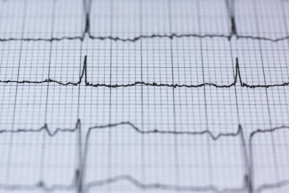
The Science: What Skin Impedance Is and Why It Matters
The ECG signal originates inside the heart and travels outward through the body to the skin’s surface, where it is detected by electrodes. But the skin itself is not a good conductor. The outermost layer — the stratum corneum — is composed of dead, keratinized cells that act as a natural electrical insulator. Oils, sweat residue, lotions, and dead cell buildup further increase the barrier.
This barrier is measured as skin impedance (or skin resistance) — the opposition the skin presents to the flow of electrical signals. Impedance is measured in ohms (Ω), and for ECG purposes, it is typically expressed in kilohms (kΩ). The higher the impedance, the harder it is for the ECG signal to reach the electrode — and the more likely the tracing will be contaminated by noise, baseline wander, and artifact.
Here is why this matters clinically: ECG monitors are designed on the assumption that all electrodes are attached to the patient’s skin with relatively low and equal impedance. When this assumption is violated — when one electrode has high impedance or when there is a significant imbalance between electrode impedances — the monitor’s common-mode rejection circuitry cannot effectively filter out electromagnetic interference (EMI). The result is artifact that can mimic arrhythmias, distort ST-segment morphology, or generate false alarms (Biomedical Instrumentation & Technology, AAMI).
354 kΩ
Average skin impedance measured before any skin preparation. At this level, the skin presents a massive barrier to the ECG signal. (Oster, Biomedical Instrumentation & Technology, 2000; Solventum/3M white paper)
20 kΩ
Average skin impedance after mild abrasion with a trace prep pad — a 94% reduction. The ECG signal can now pass through the skin with dramatically less resistance. (Same study, Oster 2000)
2.5 kΩ
Skin impedance after abrasion in a second study — down from 88.7 kΩ without prep. That is a 97% reduction, making it roughly 35 times easier for the ECG signal to reach the electrode. (Jonasson, 2007; cited in Solventum/3M white paper)
To put this in perspective: without skin prep, the ECG signal must fight through a barrier equivalent to leaving a thick layer of insulating tape between the electrode and the skin. After just a few seconds of abrasion, that barrier is essentially removed. The signal arrives at the electrode cleaner, stronger, and with far less noise.
THE KEY PRINCIPLE: ECG monitor filters are designed to work when all electrodes have low, balanced impedance. When impedance is high or imbalanced between electrodes, the monitor cannot distinguish between real cardiac signal and external interference. This means skin prep doesn’t just make the tracing look cleaner — it enables the monitor’s own technology to function as designed.
What Happens When You Skip Skin Prep
When skin preparation is skipped, the consequences are predictable and well-documented:
Baseline wander. High impedance allows low-frequency noise to contaminate the tracing, producing a slow, undulating baseline that makes ST-segment analysis unreliable. In a STEMI evaluation, even subtle baseline wander can obscure the 1–2mm of ST-segment elevation that defines the diagnosis.
High-frequency artifact. Muscle tremor, 60Hz powerline interference, and electromagnetic noise from nearby equipment are amplified when impedance is high or imbalanced. This artifact can mimic atrial fibrillation, ventricular tachycardia, or other arrhythmias, leading to false alarms and clinical distraction.
False alarms. A Solventum/3M white paper on ECG monitoring reported that nurses can be prompted by as many as 700 alarms per day, of which 80–99% are false or clinically insignificant. The FDA reported 566 deaths related to monitor alarms from 2005 to 2008 — many attributable to alarm fatigue, a condition in which caregivers become desensitized to constant false alerts. Poor skin prep is a primary contributor to the false alarm burden.
Repeat ECGs. When the first ECG is uninterpretable due to artifact, the provider must re-acquire — adding 3–5 minutes, consuming additional supplies, and delaying clinical decision-making. In a time-sensitive condition like STEMI, where every minute of delay increases mortality risk (De Luca et al., Circulation, AHA, 2004), a preventable re-acquisition is not just an inconvenience. It is a clinical risk.
Electrode detachment. Oils and moisture on unprepared skin reduce electrode adhesion. In the prehospital environment, where vehicle motion and patient diaphoresis are constant challenges, electrodes placed on unprepared skin are far more likely to peel, shift, or lose contact — degrading signal quality progressively throughout transport.
86%
of clinicians surveyed reported experiencing unacceptable ECG trace quality (52% “often,” 34% “occasionally”). (AAMI Annual Meeting / AACN NTI survey, cited in Oster 2000)
17%
of survey participants had a protocol in place that required skin preparation prior to electrode placement. The vast majority had no formal skin prep requirement. (Same survey)
How to Prep: A Quick, Evidence-Based Protocol
Proper skin preparation is fast, inexpensive, and dramatically effective. Here is the evidence-based approach:
- Step 1: Expose and inspect the skin. Remove clothing covering the electrode sites. Visually inspect for excessive hair, moisture, lotions, or skin conditions that may affect adhesion.
- Step 2: Remove excess hair if necessary. If hair is dense enough to prevent electrode contact, clip (do not shave) the hair at the electrode site. Shaving can cause micro-abrasions that introduce artifact from skin irritation.
- Step 3: Abrade lightly. Using a trace prep pad (fine abrasive), a dry 4x4 gauze, or a rough cloth, rub the electrode site firmly with 5–10 strokes until the skin appears slightly pink. This removes the stratum corneum — the dead cell layer that creates the impedance barrier. Philips application notes confirm that just one stroke of fine abrasive material can significantly reduce skin resistance (Medina et al., Heart & Lung, 1989).
- Step 4: Clean with alcohol if needed. Wipe the area with an alcohol pad to remove oils and residue, then allow it to dry completely before applying the electrode. Note: if you abrade first and then clean with alcohol, the impedance remains low. If you only use alcohol without abrasion, the benefit is minimal because the dead cell layer remains intact.
- Step 5: Apply the electrode immediately. Do not pre-attach electrodes to cables and leave them exposed — the conductive gel will dry out. Open electrode packaging just before use, press the electrode firmly over the center of the prepared site to ensure good gel-to-skin contact, and connect the cable.
PRO TIP: The entire skin prep process takes 5–10 seconds per electrode site. For a standard 10-electrode 12-lead ECG, that’s roughly 60–90 additional seconds of total scene time. In exchange, you get a tracing that is up to 97% more conductive, dramatically cleaner, and far less likely to require a repeat acquisition that costs you more time than the prep ever would.
Skin Prep and the EXG™: Better Signal, Faster Acquisition, Fewer Repeats
All ECG electrode systems — including the EXG™ Wearable ECG Platform from C-Booth Innovations — benefit from proper skin preparation. The EXG FAQ explicitly states: “All electrodes recommend skin prep to improve the fidelity of ECG tracings, including EXG. Skin prep can be easily performed with EXG prior to or during application without increasing FMC-to-electrode or scene time.”
But the EXG’s design amplifies the benefits of skin prep in ways that traditional individual electrodes cannot:
- • 20–30 second application. CBI’s clinical validation studies show that the EXG is applied in 20–30 seconds — at least 2 minutes faster than traditional lead-by-lead placement. This speed advantage means that even with skin prep added, total FMC-to-ECG time is shorter than traditional placement without skin prep.
- • Uninterpretable ECGs dropped from 7% to 3%. When an EMS system introduced the EXG along with proper patient positioning and skin prep protocols, the rate of uninterpretable ECGs was cut by more than half, and overall ECG quality improved. (CBI FAQ, citing clinical validation data.)
- • FMC-to-ECG decreased by 1 minute. Even with the addition of skin prep and patient positioning steps, the net effect was a 1-minute reduction in time to first ECG — because the EXG’s rapid deployment more than compensated for the additional prep time.
- • Improved skin impedance for high-fidelity signals. CBI reports that the EXG provides improved skin impedance characteristics compared to traditional electrodes, contributing to higher signal fidelity. Combined with proper skin prep, this means the ECG signal travels through the lowest possible impedance path from heart to monitor.
- • Validated for 3-day wear. The EXG is tested for up to 72-hour continuous wear, making it suitable for inpatient monitoring as well as prehospital and ED use. Skin prep at the time of initial application ensures that impedance remains low throughout the wear period, maintaining signal quality over days rather than degrading over hours as traditional electrode gel dries out.
- • 100% improvement in electrode placement accuracy. CBI’s data shows the EXG’s anatomically guided design ensures proper placement every time — eliminating the most common source of ECG acquisition error (precordial misplacement) while skin prep eliminates the most common source of signal degradation (high impedance).
THE COMBINED EFFECT: Proper skin prep removes the impedance barrier. Proper electrode placement ensures the signal is captured from the correct anatomical positions. The EXG delivers both — correct placement by design, combined with a system that integrates skin prep into the workflow without adding time. The result is diagnostic-quality ECG data from the first tracing, with fewer repeats, fewer artifacts, and fewer false alarms.
The Bottom Line: 10 Seconds That Protect Your Patient and Your Tracing
Skin preparation is not glamorous. It doesn’t appear on competency exams. It’s rarely emphasized in refresher training. And in the time pressure of an emergency, it’s the step most likely to be skipped.
But the science is unambiguous: unprepared skin can present an impedance barrier of 354 kΩ or more — a barrier that degrades every ECG signal passing through it. A few seconds of gentle abrasion can reduce that barrier by 94–97%, transforming a noisy, artifact-prone tracing into a clean, diagnostic-quality recording that clinicians can trust.
When you combine proper skin prep with a system like the EXG that ensures anatomically correct electrode placement in 20–30 seconds, the result is the best possible ECG data in the shortest possible time. Fewer repeat ECGs. Fewer false alarms. Fewer artifacts mimicking pathology or hiding it. More confidence in every clinical decision that follows.
Every patient deserves a tracing that accurately represents what their heart is doing. The foundation of that tracing is the connection between the electrode and the skin. Take the 10 seconds. Prep the skin. It is the simplest thing you can do to make every ECG you acquire better.
References
- Oster CD. Improving ECG trace quality. Biomedical Instrumentation & Technology. 2000;34(3):219–222. (354 kΩ average impedance pre-prep, 20 kΩ post-prep = 94% reduction.)
- Jonasson L. A prospective study on the relevance of skin preparation for noise, impedance and ECG intervals among healthy males. 2007. (88.7 kΩ pre-prep, 2.5 kΩ post-abrasion = 97% reduction.)
- Solventum (formerly 3M). Proper Skin Preparation Improves Trace Quality and Reduces ECG Monitoring Alarms. White paper. (86% of clinicians report unacceptable trace quality; 17% had skin prep protocols.)
- Medina V, Clochesy JM, Omery A. Comparison of electrode site preparation techniques. Heart & Lung: Journal of Critical Care. 1989;18(5):456–460.
- Adams-Hamoda MG, Adams H, Caldwell MA, Stotts NA, Drew BJ. Factors to consider when analyzing 12-lead electrocardiograms for evidence of acute myocardial ischemia. American Journal of Critical Care. 2003;12:9–18.
- Biomedical Instrumentation & Technology (AAMI). Electrocardiogram Interference: A Thing of the Past? (Impedance imbalance negates common-mode rejection.)
- Philips Healthcare. Improving ECG Quality — Application Note. (334 kΩ reduction with skin abrasion; stratum corneum removal protocol.)
- C-Booth Innovations. EXG™ Frequently Asked Questions. cboothinnovations.com/faq. (20–30 second application; 7% to 3% uninterpretable reduction; FMC-to-ECG decreased by 1 minute; improved skin impedance.)
- De Luca G, et al. Time delay to treatment and mortality in primary angioplasty: every minute of delay counts. Circulation. 2004;109:1223–1225.
- Gregory P, Kilner T, Lodge S, Paget S. Accuracy of ECG chest electrode placements by paramedics. British Paramedic Journal. 2021;6(1):8–14.
- FDA. Reported 566 deaths related to monitor alarms, 2005–2008. (Cited in Solventum/3M white paper.)
About C-Booth Innovations
C-Booth Innovations is pioneering precision in cardiac monitoring with the EXG™ Wearable ECG Platform — founded by two emergency room physicians who believe every patient deserves diagnostic-quality ECG data from the first tracing. The EXG is FDA registered, ISO certified, AHA-compliant, and universally compatible with existing cardiac monitors. To learn more, visit cboothinnovations.com or call (760) 800-2109.
© 2026 C-Booth Innovations. All rights reserved. | 5835 Avenida Encinas, Suite 118, Carlsbad, CA 92008


Ready to Improve ECG Accuracy?
See how the EXG platform can standardize electrode placement across your organization.